Research
Research
How the brain responds to the things we implant in it, and whether we can talk it down.
The short version
A brain-computer interface reads the electrical chatter of neurons, and sometimes writes to them, through electrodes thinner than a hair.A brain-computer interface (BCI) turns neural activity into commands for a computer or device, or sends signals the other way. They work remarkably well at first. Over months, the signal they record tends to get weaker and noisier.
One reason is the brain’s own response. To the brain, an electrode is a foreign object that arrived by injury, and it reacts to it the way it reacts to other injuries. I designed and ran a study on that reaction, and it has since been published.
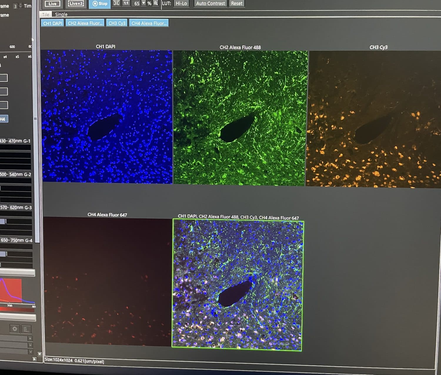
the tissue
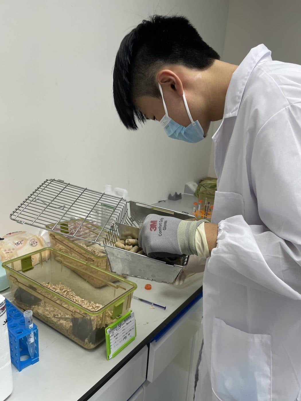
in the lab
Drag to move the photo, or use the arrow keys. Press Enter to open it.
Why implants go quiet
Putting an electrode in damages some tissue, and the device stays behind as something the body can’t clear. The brain’s resident immune cells, the microglia, move toward it. Astrocytes, the support cells that fill much of the space between neurons, join them.Microglia are the brain’s resident immune cells. Astrocytes are star-shaped support cells that, among other jobs, form scar tissue after an injury. Over weeks the cells wall the electrode off, and the neurons that had been close enough to hear can fall silent or die back. A recording site surrounded by that wall hears less.
I should say plainly that this is one part of a bigger picture. Materials, mechanics and the long-term stability of the insulation matter too, and no single fix will settle it.
How I got into it
I was pulled in by what these devices make possible. A person paralyzed from the shoulders down can move a cursor with their thoughts, answer email, and play a video game.I mean Noland Arbaugh, the first person to receive a Neuralink implant. Weeks after his surgery, many of the implant’s threads pulled back from the tissue and the signal dropped, a reminder that implants can fail in more than one way. Other interfaces drive robotic arms, aim to restore vision, or help people who can’t speak communicate again.
Then I learned that these devices can lose signal over time, and that the brain’s reaction to them is one reason why. After months of reading, one protein kept turning up: galectin-3, which activated microglia release and which drives inflammation. In studies of traumatic brain injury it rises within the first day, and a neutralizing antibody against it reduced key inflammatory signals and protected neurons.The signals were cytokines, the proteins immune cells use to call for more inflammation: IL-1β, IL-6 and TNF-α.
An implanted electrode is its own small injury. So the question wrote itself: if blocking galectin-3 helps after a head injury, might it help around an implant?
The study
I designed it myself, modeling the methods on an earlier paper that counted neurons around chronically implanted electrodes: “Neuronal cell loss accompanies the brain tissue response to chronically implanted silicon microelectrode arrays.” Mouse brains are too small for a real BCI, so a thin platinum wire stood in for the implant. The anti-Galectin-3 antibody is an investigational antibody developed by Suninflam, the biotech company where I did the work.
Mice with the wire were dosed twice a week for four weeks. I tracked motor function with the Rotarod, then sectioned the brain tissue and stained it to look at activated microglia around the wire.The Rotarod is a rotating rod. A mouse has to keep walking to stay on it, so time on the rod is a simple measure of motor function. Immunohistochemistry uses antibodies carrying a tag to light up specific cells in a slice of tissue. I analyzed the results in GraphPad Prism.
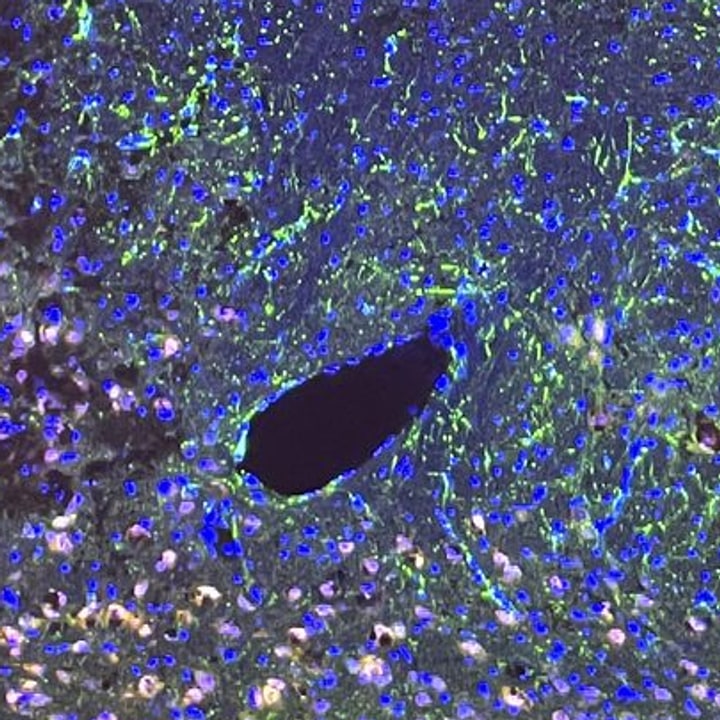
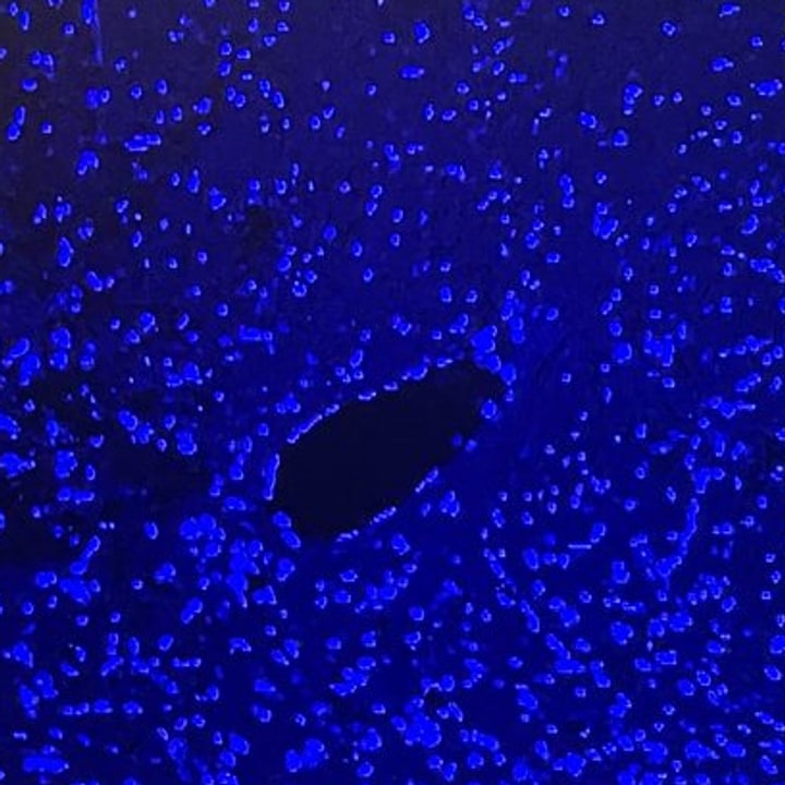
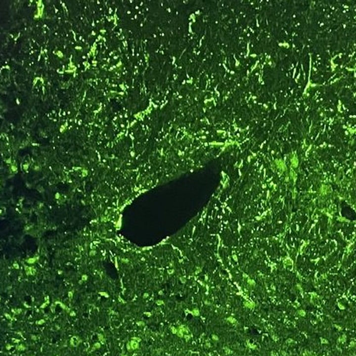
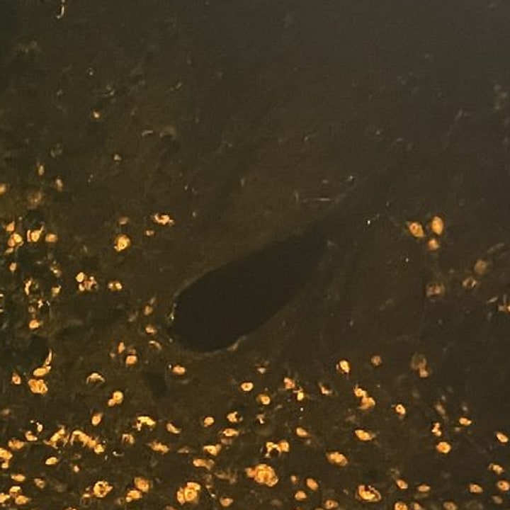

Press plus and minus to zoom, the arrow keys to pan, and zero to reset.
The four channels laid over one another. Where stains overlap, their colours mix.
What it showed
Treated mice had about 60% fewer activated microglia around the wire (p < 0.0001). The treatment group’s time on the Rotarod rose from 20 seconds to 113 seconds (p < 0.01). In plain terms: in this mouse model, the antibody was associated with a smaller immune response around the implant and with better motor outcomes.
What it can’t tell us
This was a small mouse study, with a wire standing in for a real implant. So it can show that the immune response was reduced, but it can’t show whether a real BCI would record better, which is the question that matters. It also says nothing yet about people.
Who did what
I read the literature, came up with the hypothesis, and designed the study, and I spent about six months refining the plan before I started. During a two-month summer internship I did the lab work: cutting wire, dosing, Rotarod testing, sectioning, staining, and imaging. I then spent a couple more months analyzing the data and writing it up, about ten months from idea to report.
My mentor, Dr. Suhail Rasool, trained me to handle the mice, give injections safely, section tissue on the microtome, and run and analyze the Rotarod. For safety and ethical reasons he performed the steps I didn’t: inserting the platinum wire and euthanizing the mice. The company provided the lab, equipment and animals. I wasn’t paid.
I also presented the work to my school’s Biochemistry Club, and used it to teach other students how to run a study and write up findings.
What’s next
The obvious next step is a larger animal, where a real BCI can be used and signal quality can be compared before and after treatment. I’d like to work with research groups and companies that build more advanced interfaces, and eventually build a path from mouse studies toward human ones. That is a long road, and this is its first step.
What I’m still curious about
What hooks me is that this problem sits between fields. An interface that lasts is partly biology (calm the reaction), partly materials and mechanics (be gentler to begin with), and partly signal processing (cope with a connection that keeps changing). I would like to work in the places where those have to agree with one another.
Recognition
In 2026 I received the American Academy of Neurology’s Neuroscience Research Prize, one of three high school recipients nationwide, and I presented the work at the AAN Annual Meeting in Chicago.

the three recipients
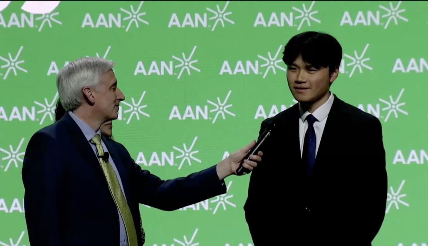
on stage

Chicago
Drag to move the photo, or use the arrow keys. Press Enter to open it.
Publications
- Sun JH, Zhao J, Sun Y, Zheng C, Zhou M, Sun D, et al. “Suppression of neuroinflammatory response to platinum wire implanted in mouse brain by monoclonal antibody SIF001 against Galectin-3.” American Journal of Biomedical Science & Research, 2025; 27(5). DOI: 10.34297/AJBSR.2025.27.003591. First author.
- Rasool S, Sun JH, Zhou M, Sun Y, Zheng C, et al. “Modulation of Neuroinflammatory Responses to Neural Implants.” Journal of Neurology and Neurological Disorders, 2025; 11(1): 102. Second author.
Read more
- The NeuroView lecture on BCIs and the foreign body response explains this same problem from the beginning.
- A Beginner’s Guide to BCIs, the article I wrote for NeuroView.